Ki-67 Rabbit mAb(ABT104R) (Ready to Use)
- Catalog No.:YM7002R
- Applications:IHC
- Reactivity:Human
- Target:
- Ki-67
- Gene Name:
- MKI67
- Protein Name:
- Ki 67
- Human Gene Id:
- 4288
- Human Swiss Prot No:
- P46013
- Immunogen:
- Synthesized peptide derived from human Ki-67
- Specificity:
- The antibody can specifically recognize human Ki-67 protein.
- Formulation:
- Liquid in PBS containing 50% glycerol, 0.05% proclin 300
- Source:
- Monoclonal, Rabbit,IgG1, Kappa
- Dilution:
- Ready to use for IHC
- Purification:
- Recombinant Expression and Affinity purified
- Storage Stability:
- 2°C to 8°C/1 year
- Other Name:
- MKI67;Antigen KI-67
- Background:
- This gene encodes a nuclear protein that is associated with and may be necessary for cellular proliferation. Alternatively spliced transcript variants have been described. A related pseudogene exists on chromosome X. [provided by RefSeq, Mar 2009],
- Function:
- developmental stage:Expression of this antigen occurs preferentially during late G1, S, G2 and M phases of the cell cycle, while in cells in G0 phase the antigen cannot be detected.,function:Thought to be required for maintaining cell proliferation.,online information:Ki-67 entry,similarity:Contains 1 FHA domain.,subcellular location:Predominantly localized in the G1 phase in the perinucleolar region, in the later phases it is also detected throughout the nuclear interior, being predominantly localized in the nuclear matrix. In mitosis, it is present on all chromosomes.,subunit:Interacts with KIF15. Binds through the FHA domain to MKI67IP.,
- Subcellular Location:
- Nuclear
- Expression:
- Nuclear
- June 19-2018
- WESTERN IMMUNOBLOTTING PROTOCOL
- June 19-2018
- IMMUNOHISTOCHEMISTRY-PARAFFIN PROTOCOL
- June 19-2018
- IMMUNOFLUORESCENCE PROTOCOL
- September 08-2020
- FLOW-CYTOMEYRT-PROTOCOL
- May 20-2022
- Cell-Based ELISA│解您多样本WB检测之困扰
- July 13-2018
- CELL-BASED-ELISA-PROTOCOL-FOR-ACETYL-PROTEIN
- July 13-2018
- CELL-BASED-ELISA-PROTOCOL-FOR-PHOSPHO-PROTEIN
- July 13-2018
- Antibody-FAQs
- Products Images

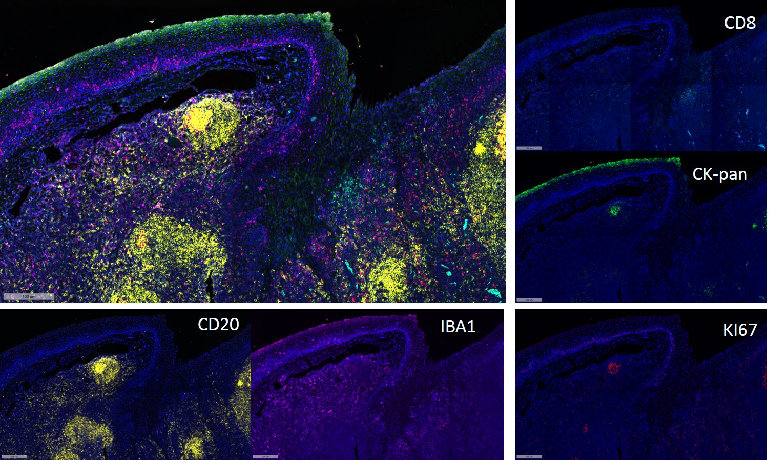
- Fluorescence multiplex immunohistochemical analysis of Human tonsil tissue (formalin-fixed paraffin-embedded section). The immunostaining was performed by Pentuple-Fluorescence kit (RS0038, Immunoway). CK-pan mouse mAb(YM6815 Immunoway) green, Ki-67 rabbit mAb(YM7002 Immunoway) red, Iba 1 mouse mAb(YM4765 Immunoway) purple,CD8 a mouse mAb(YM4815 Immunoway) cyan, CD20 mouse mAb(YM4814 Immunoway) yellow, The section was incubated in 5 rounds of staining; sequentially for Anti-antibodies; each using a separate fluorescent tyramide signal amplification system. EDTA based antigen retrieval (Immunoway YS0004, pH 9.0, 20 minutes) was used in between rounds of tyramide signal amplification to remove the antibody from the previous round, to avoid any cross-reactivity. DAPI (dark blue) was used as a nuclear counter stain. Microscopy and pseudocoloring of individual dyes was performed using a Slideviewer Imaging System (Excilone).


