p21 (PTR2559) mouse mAb
- Catalog No.:YM3802
- Applications:IHC;WB;IF;ELISA
- Reactivity:Human;Mouse;
- Target:
- P21
- Gene Name:
- CDKN1A CAP20 CDKN1 CIP1 MDA6 PIC1 SDI1 WAF1
- Protein Name:
- p21
- Human Gene Id:
- 1026
- Human Swiss Prot No:
- P38936
- Mouse Gene Id:
- 12575
- Mouse Swiss Prot No:
- P39689
- Immunogen:
- Synthesized peptide derived from human p21 AA range: 1-100
- Specificity:
- This antibody detects endogenous levels of p21 protein.
- Formulation:
- PBS, 50% glycerol, 0.05% Proclin 300, 0.05%BSA
- Source:
- Mouse, Monoclonal/IgG1, kappa
- Dilution:
- IHC 1:200-1000. WB 1:500-2000. IF 1:100-500. ELISA 1:1000-5000
- Purification:
- Protein G
- Concentration:
- 1 mg/ml
- Storage Stability:
- -15°C to -25°C/1 year(Do not lower than -25°C)
- Other Name:
- Cyclin-dependent kinase inhibitor 1 ;CDK-interacting protein 1;Melanoma differentiation-associated protein 6;MDA-6;p21;
- Molecular Weight(Da):
- 21kD
- Observed Band(KD):
- 21kD
- Function:
- function:May be the important intermediate by which p53 mediates its role as an inhibitor of cellular proliferation in response to DNA damage. Binds to and inhibits cyclin-dependent kinase activity, preventing phosphorylation of critical cyclin-dependent kinase substrates and blocking cell cycle progression.,induction:By p53, mezerein (antileukemic compound) and interferon beta.,PTM:Phosphorylation of Thr-145 by Akt or of Ser-146 by PKC impairs binding to PCNA.,similarity:Belongs to the CDI family.,tissue specificity:Expressed in all adult human tissues, with 5-fold lower levels observed in the brain.,
- Subcellular Location:
- Cytoplasmic, Nuclear
- Expression:
- Expressed in all adult tissues, with 5-fold lower levels observed in the brain.
- June 19-2018
- WESTERN IMMUNOBLOTTING PROTOCOL
- June 19-2018
- IMMUNOHISTOCHEMISTRY-PARAFFIN PROTOCOL
- June 19-2018
- IMMUNOFLUORESCENCE PROTOCOL
- September 08-2020
- FLOW-CYTOMEYRT-PROTOCOL
- May 20-2022
- Cell-Based ELISA│解您多样本WB检测之困扰
- July 13-2018
- CELL-BASED-ELISA-PROTOCOL-FOR-ACETYL-PROTEIN
- July 13-2018
- CELL-BASED-ELISA-PROTOCOL-FOR-PHOSPHO-PROTEIN
- July 13-2018
- Antibody-FAQs
- Products Images
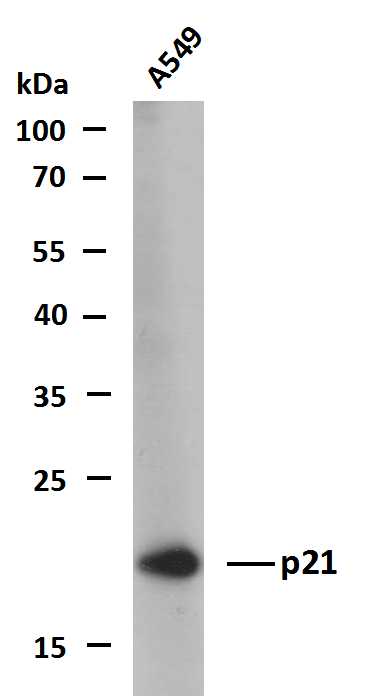
- Whole cell lysates of A549 were separated by 12% SDS-PAGE, and the membrane was blotted with anti-p21(PTR2559) antibody. The HRP-conjugated Goat anti-Mouse IgG(H + L) antibody was used to detect the antibody. Lane 1: A549
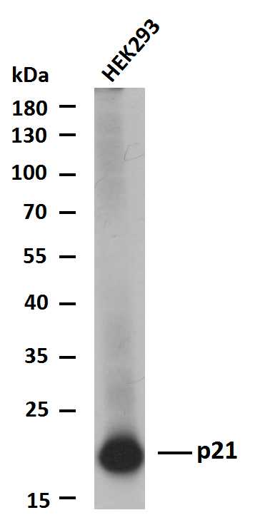
- Whole cell lysates of HEK293 were separated by 12% SDS-PAGE, and the membrane was blotted with anti-p21(PTR2559) antibody. The HRP-conjugated Goat anti-Mouse IgG(H + L) antibody was used to detect the antibody. Lane 1: HEK293

- Whole cell lysates of MCF7 were separated by 12% SDS-PAGE, and the membrane was blotted with anti-p21(PTR2559) antibody. The HRP-conjugated Goat anti-Mouse IgG(H + L) antibody was used to detect the antibody. Lane 1: MCF7


